How Does Ultrasound Work?
sophiaisabelle October 12, 2021An ultrasound machine uses ultrasound waves to show pictures of what’s going on inside the body. A transducer instrument sends out a high-frequency sound that humans can’t hear and then records echoes as the sound waves bounce back and determine the size, shape, and consistency of soft tissues and organs.
How do ultrasounds work exactly?
The ultrasound machine transmits high-frequency waves (1 to 5 megahertz). Ultrasound machine Sound waves get reflected back to the probe & reach another boundary, and get reflected in the image.
What do they use to do to an ultrasound?
Sound waves get reflected back to the search, while some travel on further until they reach another edge and get remembered. More items…
How is ultrasound different to sound?
Ultrasound machines derived the term of sound or as the subject.
The difference between ultrasound and sound is that a (physics) sound with a frequency is more significant & the limit of human hearing is approximately 20 kilohertz.

What are the uses of ultrasound?
The ultrasound machine is using to looking babies in the mother’s womb.
Ultrasound machines can be used to work out how old the baby is in the womb.
Baby (male or female) checks for a heartbeat, average fetal growth, and any abnormalities.
What is a sonogram? Know its types, procedures, and uses?
Know about sonogram, their types, procedures, and uses. It is an imaging technique performed by doctors to detect any structural abnormalities in the body. A sonogram is commonly used to see swelling, growth, tumors, developing fetus in the womb, or the presence of any abnormalities.
Ultrasound Machine worker called?
A person who performs an ultrasound scan is called a sonographer.
And the images are interpreted by radiologists, cardiologists, or other specialists.
How are ultrasounds used for the heart?
Common Ultrasound type of heart is non-invasive and very easy on the patient.
A trained cardiac sonographer uses the gel to probe-transducer. And it allows reflected sound waves to provide a live picture of your heart.
Is an ultrasound considered nuclear medicine?
Nuclear medicine in diagnosis. Other types of imaging involved in nuclear medicine include targeted molecular ultrasound, which helps detect different kinds of cancer and highlight blood flow, and magnetic resonance sonography, which has a role in diagnosing cancer and metabolic disorders.
What are precautions for an ultrasound?
Precautions Needed While Undergoing an Ultrasound: Although ultrasound is a painless and harmless process, certain precautions are required to ensure better safety and health of the mother and child. These include: Wear loose clothes so that the doctor can easily access your belly.
What can you drink before an ultrasound?
Drink of water before the ultrasound is good before an ultrasound.
An accurate picture of your baby can be easily obtained image if you take water before ultrasound.
Drinking-Water tries at least 24 oz.
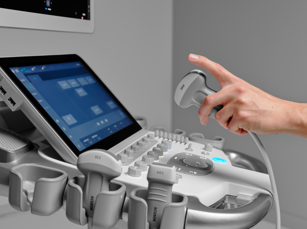
TYPES OF ULTRASOUND PROBES
There are three most common ultrasound transducers (probes): convex, linear, and phased array.
Although there exist more classes in the market, I will delve deeper into three common types of ultrasound probes.
THE CONVEX TRANSDUCERS (PROBES)
Convex ultrasound (probe), also known as a curved transducer, has its frequency, and applications depend on whether the product is meant for 2 to 3 dimensions. A comprehensive footprint product and the central frequency ranging between 2.5MHz and 7.5MHz. It is mainly used for abdominal,
PHASED ARRAY PROBES
Ultrasound Machine phased array transducers are named after the way their piezoelectric crystals are arranged. PHASED ARRAY PROBES frequency 2MHz and &.5MHz. The beam point is mainly used in cardiac examinations, including trans-esophageal tests and abdominal and brain exams.
LINEAR PROBE
Transducers’ footprint, frequency, and application often depend on whether the product is 2D or 3D imaging. Ultrasound Machine LINEAR PROBE frequency that ranges 7.5MHz to 11MHz. Generally used in breast, thyroid, and arterial carotids of vascular application.
TVS ,Transvaginal ultrasound probe
Vaginal ultrasonography is medical ultrasonography that applies an ultrasound transducer (or “probe”) in the vagina. And visualize organs within the pelvic cavity.
It is also called transvaginal, ultrasonography.
Why would a transvaginal ultrasound be painful?
Regular: It is not uncommon to have cramps after the transvaginal US or pelvic exam since a foreign body – ultrasound probe or gloved hand often causes a release of prostaglandins leading to uterine cramping.
OTHER TYPES OF ULTRASOUND PROBES
PENCIL TRANSDUCERS
This transducer is also called the CW Doppler probe & it is using for memories blood flow.
Its l frequency, typically 2MHz to 8MHz.
ENDO-CAVITARY PROBES
These are ultrasound probes that allow internal examination of a patient; they are designed to fit into the human openings like the rectum and vagina. The frequencies range of ENDO-CAVITARY PROBES 3.5MHz to 11.5MHz.
ULTRASOUND Machine BENEFIT
Ultrasound machines waves can produce high-quality images without emitting harmful radiations like x-ray, and the likes do.
They are even safe for pregnancy. The ultrasound machines are portable and inexpensive nowadays; you can hardly find a local doctor nearby.
ULTRASOUND PROBES repairing
Ultrasound Machine probe repairs are inevitable because probe repair costs are sometimes a tiny fraction of the actual price.
What are the advantages of an ultrasound?
Advantages of Using Ultrasound for Diagnostic Imaging. Ultrasound is safe, nonionizing, effective, affordable, easy to learn, and simple to interpret. It provides immediate results, allows for live assessment of organs and pain points, and can easily be shared with colleagues.
What are the disadvantages of ultrasound?
It has poor penetration through bone or air. Moreover, it has limited penetration in obese patients. These are the disadvantages in the imaging domain.
Should ultrasound be painful?
No, most pelvic ultrasounds are not painful. However, they can be painful when they are used in certain conditions. If your pelvic muscles are in spasm, this may cause pain.


